Perfusion
Background
Perfusion MRI typically refers to MRI-based measurements of microcirculatory parameters such as blood flow, blood volume and blood mean transit time. Imaging of perfusion-related parameters is of relevance in, for example, studies of dementia and trauma, assessment of the ischemic penumbra in acute ischemic stroke (in combination with diffusion MRI) to facilitate treatment decisions, investigations of intracranial vascular malformation in connection with neurointerventional procedures, preoperative classification and grading of brain tumours and monitoring of various kinds of therapy.
Specific Work
The perfusion MRI research within our group is primarily focused on development and validation of MRI pulse sequences and post-processing tools for assessment of perfusion-related parameters and on improved estimation of cerebral blood flow in absolute terms.
The most common method for brain perfusion imaging in clinical environments is dynamic susceptibility contrast MRI (DSC-MRI). This approach is based on dynamic monitoring of tracer concentration, during the first passage, in an artery (for registration of the arterial input function (AIF)) and in the tissue of interest, following rapid intravenous injection of a gadolinium contrast agent. The concept is based on the theory of intravascular tracers and provides parametric maps of regional cerebral blood flow (CBF), cerebral blood volume (CBV) and mean transit time (MTT).
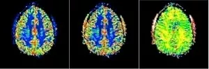
DSC-MRI at 7T: Parametric maps of CBF, CBV and MTT
Arterial spin labelling (ASL) is a completely non-invasive technique for CBF assessment, based on the use of magnetically labelled arterial water as an endogenous, diffusible tracer. Magnetic labelling or tagging of blood water spins is accomplished by inversion of the spin population in a tissue-feeding artery. The labelled water protons are then transported with the blood to the brain tissue, where the labelled spins change the net magnetization in proportion to the tissue blood flow.
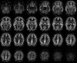
High-quality perfusion maps at 3T acquired in less than 5 minutes using pseudo-continuous arterial spin labeling (PCASL) with segmented 3D-GRASE readout.
Another non-invasive approach to determine perfusion-related parameters, primarily the so-called perfusion fraction (related to blood volume), is intravoxel incoherent motion (IVIM) imaging, in which diffusion encoding is used to extract phase dispersion effects caused by incoherent motion of the spin population within the pseudo-randomly oriented blood capillaries of the microvasculature.
The blood supply in terms of CBF is closely related to the subsequent cerebral oxygen delivery and oxygen consumption, and a number of MRI approaches have been proposed to quantify venous oxygen saturation, oxygen extraction fraction (OEF) and cerebral metabolic rate of oxygen (CMRO2). MRI methods for OEF assessment include T2/T2* measurements, phase or magnetic susceptibility maps, and spin labelling for obtaining pure venous blood signal.
Aims
- To investigate the role of quantitative susceptibility mapping (QSM) for contrast agent quantification in perfusion imaging
- To explore new blood-volume-weighted and transit-time-related estimates to be used as a supplement to ASL-based CBF and, potentially, for DSC-MRI calibration.
- To develop post-processing and analysis tools for DSC-MRI and ASL data, for example, deconvolution algorithms.
- To estimate blood oxygen saturation levels, primarily using QSM, for assessment of oxygen extraction fraction (OEF) and cerebral metabolic rate of oxygen (CMRO2).
- To implement and establish a variety of perfusion methodologies in the clinical environment.
Representative Perfusion MRI Publications
Ahlgren A, Wirestam R, Knutsson L, Petersen ET. Improved calculation of the equilibrium magnetization of arterial blood in arterial spin labeling. Magn Reson Med. 2018 Nov;80(5):2223-2231. doi: 10.1002/mrm.27193.
Knutsson L, Xu X, Ståhlberg F, Barker PB, Lind E, Sundgren PC, van Zijl PCM, Wirestam R. Dynamic susceptibility contrast MRI at 7 T: Tail-scaling analysis and inferences about field strength dependence. Tomography. 2017 Jun;3(2):74-78. doi: 10.18383/j.tom.2017.00001.
Lind E, Knutsson L, Kämpe R, Ståhlberg F, Wirestam R. Assessment of MRI contrast agent concentration by quantitative susceptibility mapping (QSM): application to estimation of cerebral blood volume during steady state. MAGMA. 2017 Dec;30(6):555-566. doi: 10.1007/s10334-017-0637-9.
Rydhög AS, Szczepankiewicz F, Wirestam R, Ahlgren A, Westin CF, Knutsson L, Pasternak O. Separating blood and water: Perfusion and free water elimination from diffusion MRI in the human brain. Neuroimage. 2017 Aug 1;156:423-434. doi: 10.1016/j.neuroimage.2017.04.023.
For more information about our perfusion MRI research please contact:
Ronnie Wirestam, Ph.D.
Professor of Medical Radiation Physics
Dept. of Medical Radiation Physics, Lund University, Lund, Sweden
e-mail: ronnie [dot] wirestam [at] med [dot] lu [dot] se (ronnie[dot]wirestam[at]med[dot]lu[dot]se)
LU rsearch portal profile: Ronnie Wirestam
Perfusion
Group Leader
Ronnie Wirestam, Ph.D.
Professor
e-mail: ronnie [dot] wirestam [at] med [dot] lu [dot] se (ronnie[dot]wirestam[at]med[dot]lu[dot]se)
LU research portal profile: Ronnie Wirestam

Seniors
Lnda Knutsson, Ph.D.
Professor
e-mail: linda [dot] knutsson [at] med [dot] lu [dot] se (linda[dot]knutsson[at]med[dot]lu[dot]se)
LU research portal profile: Linda Knutsson

Emelie Lind, Ph.D.
e-mail: emelie [dot] lind [at] med [dot] lu [dot] se (emelie[dot]lind[at]med[dot]lu[dot]se)
LU research portal profile: Emelie Lind

Doctoral Students
Anna Lundberg, M.Sc.
e-mail: anna [dot] lundberg [at] med [dot] lu [dot] se (anna[dot]lundberg[at]med[dot]lu[dot]se)
LU research portal profile: Anna Lundberg